


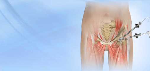

Hip Labrum Tear web based movie
The Acetabular Labrum is a ring of strong fibrocartilaginous tissue on the rim of the hip socket (acetabulum). The Labrum serves many functions: it acts as a shock absorber, lubricates the joint, and distributes pressure equally. It holds the head of the femur in place and prevents the lateral and vertical movement of the femur head within the joint. It also deepens the acetabular cavity and offers stability against femoral head translation.
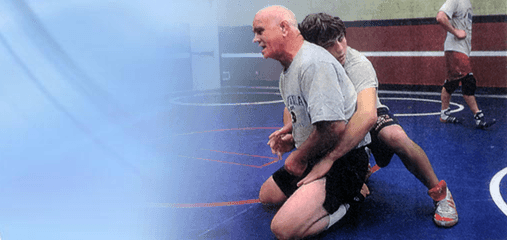
An acetabular labrum tear (or hip labral tear) may be caused by trauma, femoroacetabular impingement (FAI), hip hypermobility, dysplasia, and degeneration. It is common in athletes playing sports such as ice hockey, soccer, golf and ballet. Structural abnormalities may also cause hip labral tears.
Patients with acetabular labrum tears frequently have pain in the groin region, but may also in the outside or back of the hip. Hip Labral Tears may cause clicking, catching and/or locking of the joint and restricted range of motion. Patients may also experience dull pain on movement of hip joint that may or may not subside with rest. Hip labral tear is often diagnosed by symptoms, history, physical examination and radiological techniques. Regular x-rays of the hip help to determine whether the hip joint has normal anatomy or evidence of impingement or arthritis.
Because the acetabular labrum is made of soft tissue, it usually cannot be seen on regular x-rays. For this reason, patients who have symptoms of an acetabular labrum tear are usually sent for a Magnetic Resonance Imaging Arthrogram, also known as “MRI arthrography” or an “MRI with contrast.” In this test, the radiologist or surgeon injects the hip joint with a clear liquid that shows up on the MRI images (“contrast”). The contrast liquid can find its way into the torn portion of the labrum. A “positive” MRI with contrast helps confirm the presence of the labrum tear and helps identify where the tear is located.
However, it should be noted that hip labrum tears are difficult to image. In other words, the MRI with contrast may not show a tear even though it is present. This is referred to as a “false negative.” Dr. Grimes has been performing hip arthroscopy for over 20 years. During that time, approximately 50% of patients who were arthroscopically confirmed to have a labrum tear had a negative hip MRI arthrogram.
Your doctor may start with conservative treatment prescribing non-steroidal anti-inflammatory drugs and advising you to rest. These methods may offer symptomatic relief while surgery is required to treat the torn labrum. Your doctor may perform arthroscopic surgery using fiber-optic camera and surgical instruments through the smaller incisions. Depending on the type and severity of tear, the damaged or torn part of the labrum may be removed or reattached.